A recent clinical trial has found that PSMA PET/CT and conventional bone scans have identical detection rates for bone metastases in prostate cancer patients with rising PSA levels during hormone therapy. This suggests that the more accessible and cost-effective bone scans can continue to play an important role in monitoring disease progression, even as PSMA PET/CT becomes more widely available.
Comparing Two Imaging Techniques for Prostate Cancer
Prostate cancer is a complex disease, and monitoring its progression is crucial for effective treatment. When prostate cancer patients on hormone therapy (known as androgen deprivation therapy or ADT) experience a rise in their PSA levels, it’s a sign that the cancer may be spreading. At this stage, doctors need to determine the extent of the disease to guide the next steps in treatment.
Two imaging techniques have emerged as valuable tools for this task: PSMA PET/CT and bone scans. PSMA PET/CT uses a radioactive tracer that binds to a protein called prostate-specific membrane antigen (PSMA), which is often overexpressed in prostate cancer cells. This allows the scan to detect even small metastases. Bone scans, on the other hand, use a radioactive tracer that highlights areas of increased bone activity, which can indicate the presence of bone metastases.
A Head-to-Head Comparison
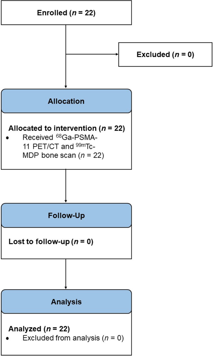
To determine which imaging technique is more effective for patients with rising PSA levels during ADT, researchers conducted a clinical trial. They enrolled 22 men with prostate cancer and had them undergo both PSMA PET/CT and bone scans. The scans were then analyzed by a team of independent radiologists to determine the presence and number of bone metastases.
The results were quite surprising: both imaging techniques had identical detection rates for bone metastases. In other words, the two scans found the same number of bone lesions in 73% of the patients. In the remaining 27% of patients, one scan detected more lesions than the other, but the difference was not statistically significant.
Implications for Clinical Practice
These findings have important implications for how prostate cancer patients are monitored during ADT. PSMA PET/CT has become increasingly popular due to its high sensitivity, but it is also more expensive and not as widely available as bone scans. The researchers suggest that in situations where cost and accessibility may be a concern, bone scans can continue to serve as a cost-effective and accessible option for restaging patients with rising PSA levels.
However, the study did find that PSMA PET/CT was better at detecting extra-osseous (non-bone) metastases. Nearly one-third of patients had additional metastases identified by PSMA PET/CT that were not visible on the conventional imaging. This highlights the value of PSMA PET/CT in providing a more comprehensive picture of disease spread.
The Road Ahead
The researchers note that this study had some limitations, such as the small sample size and the use of planar bone scans instead of the more advanced SPECT/CT scans. Further research with larger patient cohorts and longer follow-up is needed to fully understand the role of these imaging techniques in prostate cancer management.
Nonetheless, this study provides important insights into the strengths and limitations of PSMA PET/CT and bone scans. As new imaging technologies continue to emerge, clinicians will need to carefully weigh the trade-offs between cost, accessibility, and diagnostic accuracy to ensure that prostate cancer patients receive the best possible care.
Author credit: This article is based on research by John Nikitas, Andrei Gafita, Matthias R. Benz, Loïc Djaïleb, Andrea Farolfi, Masatoshi Hotta, Ida Sonni, Rejah Alano, Matthew Rettig, John Shen, Wesley Armstrong, Tristan Grogan, Sandy Liu, Johannes Czernin, Jeremie Calais.
For More Related Articles Click Here