
Navigating the intricate pathways of the human brain can be a daunting task for medical students, but a groundbreaking study has uncovered a game-changing solution. Researchers at the Radboud University Medical Center in the Netherlands have compared the effectiveness of augmented reality (AR) and traditional anatomical atlases in helping students prepare for neuroanatomy dissection sessions. The results reveal that while both methods lead to similar improvements in anatomy test scores, the use of AR technology significantly boosts students’ cognitive engagement during the preparatory phase. Augmented reality allows students to virtually interact with and manipulate 3D models of the brain, providing a more immersive and engaging learning experience. This study highlights the potential of AR to revolutionize the way we teach and learn neuroanatomy, making complex structures more accessible and captivating for the next generation of medical professionals.
Neuroanatomy Education with Augmented Reality
Understanding the intricate structure and function of the human brain is a crucial aspect of medical education, but traditional teaching methods can often fall short in effectively conveying this complex information. Anatomy lessons typically rely on a combination of textbooks, 2D diagrams, and hands-on dissection sessions, but these approaches can be limited in their ability to fully capture the three-dimensional nature of the brain.
Enter augmented reality, a technology that seamlessly integrates digital content with the physical world. By overlaying virtual 3D models of the brain onto the real-world environment, AR provides students with a more immersive and interactive learning experience. This innovative approach has the potential to revolutionize the way we teach and learn neuroanatomy.
Comparing AR and Anatomical Atlases in Neuroanatomy Preparation
To explore the effectiveness of AR in neuroanatomy education, researchers at the Radboud University Medical Center conducted a study comparing the use of an AR application and traditional anatomical atlases during student preparation for a neuroanatomy dissection session.
The study involved 28 first-year and second-year medical and biomedical science students, who were randomly divided into two groups. One group used a tablet-based AR application called “GreyMapp-AR: Brain” to complete a preparatory assignment, while the other group used the Sobotta Anatomical Atlas, a widely-used educational resource.

Both groups then participated in a body donor-based education session at the dissection room, where they worked with similar prosections of brain specimens and plastic models. Throughout the study, the researchers measured the students’ performance on anatomy tests, as well as their level of cognitive engagement during the preparatory assignments and the dissection session.
Cognitive Engagement Soars with AR
The results of the study were fascinating. While both groups showed similar improvements in their anatomy test scores, the researchers found a significant difference in the level of cognitive engagement experienced by the students.
Students who used the AR application during their preparatory assignment reported higher levels of cognitive engagement, indicating that they were more deeply involved and invested in the learning process. This suggests that the interactive and immersive nature of AR can effectively captivate students’ attention and stimulate their interest in the subject matter.

Fig. 2
Interestingly, the level of cognitive engagement was comparable between the two groups during the body donor-based education session at the dissection room. This suggests that both AR and traditional anatomical atlases can effectively prepare students for the hands-on learning experience, but the AR application may have a distinct advantage in the preparatory phase.
Implications for the Future of Neuroanatomy Education
The findings of this study have important implications for the future of neuroanatomy education. While both AR and anatomical atlases proved to be effective in helping students improve their anatomy test scores, the increased cognitive engagement observed with the AR application highlights its potential as a valuable tool in the learning process.
By leveraging the interactive and immersive capabilities of AR, educators can create a more engaging and captivating learning environment, which can ultimately lead to better retention of information and a deeper understanding of the complex structures and functions of the brain.
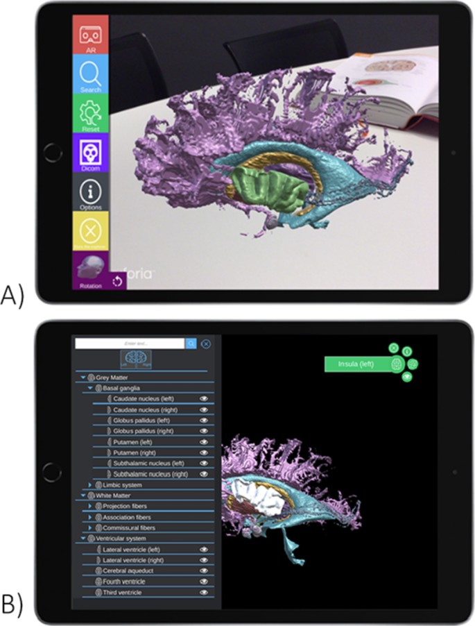
Fig. 3
Moreover, the study’s findings suggest that AR could serve as a valuable preparatory tool, helping students better prepare for and engage with hands-on dissection sessions. As medical curricula continue to evolve, with a decrease in the hours dedicated to anatomy teaching, the use of AR may become an increasingly important complement to traditional teaching methods.
the Brain’s Secrets: Future Directions and Broader Implications
While this study provides promising insights into the potential of AR in neuroanatomy education, the researchers acknowledge that further research is needed to fully understand the long-term impact of this technology. Future studies should investigate the effects of AR on students’ recall and retention of information, as well as its impact on their performance during the actual dissection sessions.
Additionally, the researchers suggest exploring the use of quantitative assessment tools, such as biosensors, to gain a deeper understanding of the cognitive processes involved when students work with AR applications. Cognitive engagement is a critical factor in the learning process, and being able to measure it more precisely could provide valuable insights into the effectiveness of different teaching methods.
Beyond the realm of neuroanatomy, the findings of this study have broader implications for the integration of AR technology in various educational settings. As medical education continues to evolve, the use of AR could extend to other areas of anatomy, physiology, and even clinical skills training, providing students with a more engaging and immersive learning experience.
As the field of augmented reality continues to advance, the potential for its application in education is vast. This study serves as a compelling example of how this innovative technology can revolutionize the way we approach complex subjects, making them more accessible and captivating for the next generation of medical professionals.
Author credit: This article is based on research by Ine Zeedzen-Scheffers, Jort Karstens, Marianne van den Hurk, Dylan Henssen, Lucas L. Boer.
For More Related Articles Click Here