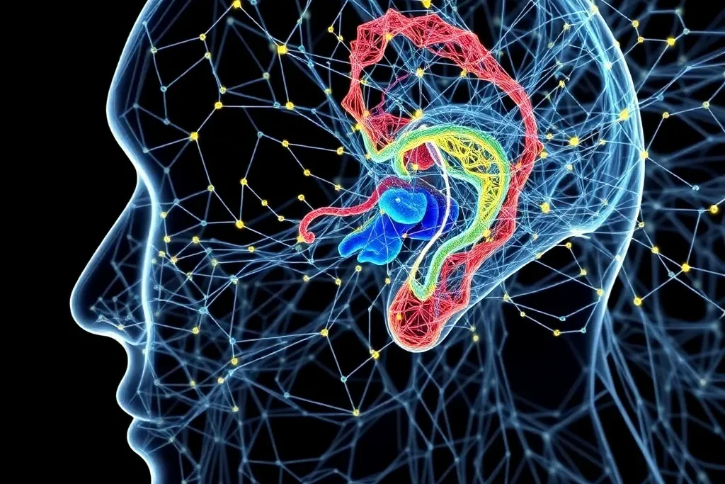
Endolymphatic hydrops (EH) is a critical condition affecting the inner ear, often linked to debilitating diseases like Ménière’s disease. Researchers have developed a groundbreaking technique using 3D neural networks to automate the analysis of EH from magnetic resonance imaging (MRI) data. This innovative approach could significantly improve the accuracy and efficiency of diagnosing inner ear disorders, ultimately enhancing patient care. The study, led by a team from Jeonbuk National University, showcases the power of advanced deep learning algorithms in revolutionizing medical imaging analysis. Endolymphatic hydrops, Ménière’s disease, magnetic resonance imaging, deep learning, neural networks.
Unraveling the Mysteries of Endolymphatic Hydrops
Endolymphatic hydrops (EH) is a critical condition affecting the inner ear, characterized by an increase in endolymphatic fluid volume. This pathological anatomical feature can lead to various inner ear illnesses, such as Ménière’s disease and fluctuating sensorineural hearing loss. Historically, the only way to confirm the presence of EH was through postmortem histological examination of temporal bone specimens. However, this invasive approach is not feasible for living patients.
The Breakthrough of Magnetic Resonance Imaging (MRI) in EH Diagnosis
The advent of magnetic resonance imaging (MRI) technology has revolutionized the way clinicians can diagnose EH. MRI techniques, such as MR cisternography (MRC) and HYDROPS-Mi2, allow for non-invasive visualization and quantification of the inner ear’s fluid spaces, including the perilymph and endolymph. This has paved the way for a deeper understanding of inner ear disorders and the potential for earlier, more accurate diagnoses.
Automating EH Analysis with 3D Neural Networks
Researchers from Jeonbuk National University have developed a groundbreaking technique to automate the analysis of EH from MRI data using 3D neural networks. Their approach involves segmenting the inner ear structures, including the cochlea, vestibule, and semicircular canals, as well as the endolymphatic fluid space, using a 3D U-Net deep learning model.

The key innovation of this study is the use of a data pair strategy, where the researchers leveraged the aligned MRC and HYDROPS-Mi2 image stacks to simultaneously segment the perilymph and endolymph fluid spaces. This end-to-end learning approach significantly improves the accuracy and efficiency of EH volume ratio calculations, which are crucial for diagnosing inner ear diseases.
Impressive Results and Clinical Implications
The researchers’ 3D segmentation model achieved impressive performance, with a Dice similarity coefficient (DSC) of 0.9574 for the total fluid space and 0.8400 for the endolymph fluid space. Moreover, the EH volume ratio calculated by the deep learning model showed high agreement with the ground truth values, as evidenced by the high intraclass correlation coefficient (ICC) of 0.951 and the Bland-Altman plot analysis.
These results demonstrate the power of 3D neural networks in accurately and efficiently quantifying EH from MRI data. This breakthrough has significant clinical implications, as it can potentially improve the diagnosis and management of inner ear disorders, such as Ménière’s disease, by providing a more objective and standardized assessment of EH.
Overcoming Challenges in Inner Ear Imaging
Segmenting the inner ear structures and the small, irregularly shaped endolymphatic fluid space is inherently challenging due to the complex anatomy and the limited size of these structures compared to the overall MRI volume. The researchers overcame these obstacles by leveraging advanced deep learning techniques and the unique data pair strategy, which allowed for simultaneous segmentation of the perilymph and endolymph fluid spaces.
This innovative approach not only improves the accuracy of EH analysis but also reduces the time and effort required by clinicians, who traditionally had to manually segment these structures, a laborious and error-prone process.
Future Directions and Broader Impact
The researchers acknowledge that their study has some limitations, such as the relatively small dataset and the need for further validation in diverse clinical settings. However, they are optimistic about the potential of their technology to be integrated into the medical decision-making process, ultimately enhancing patient care.
Beyond the immediate clinical applications, this research showcases the transformative power of 3D neural networks in the field of medical imaging analysis. The ability to automate the segmentation and quantification of complex anatomical structures, such as the inner ear, has far-reaching implications for the diagnosis and management of a wide range of medical conditions. As deep learning continues to advance, we can expect to see even more innovative solutions that revolutionize the way clinicians approach patient care.
Author credit: This article is based on research by Tae-Woong Yoo, Cha Dong Yeo, Minwoo Kim, Il-Seok Oh, Eun Jung Lee.
For More Related Articles Click Here
