When it comes to children’s eye health, persistent hyperplastic primary vitreous (PHPV) is a rare but important condition that parents and healthcare providers should know about. This congenital eye condition affects the development of blood vessels in a baby’s eye before birth, potentially leading to vision problems if not caught early.
What is PHPV?
PHPV occurs when the temporary blood vessel system that nourishes a baby’s developing eye fails to disappear as it should before birth. Think of it like scaffolding used during construction – it’s necessary during development but should be removed once the job is done. In PHPV, this “scaffolding” of blood vessels stays behind, potentially causing problems with vision.
Key Facts About PHPV:
- It typically affects only one eye (unilateral)
- It’s present from birth
- It can lead to cataracts in children
- Early detection is crucial for better outcomes
- It affects roughly 11% of children with pediatric cataracts
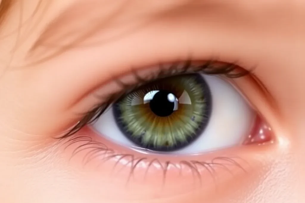
Common Signs and Symptoms:
- Clouding of the lens (cataract)
- White pupil
- Smaller than normal eye
- Vision problems
- Misalignment of the eyes
How Does PHPV Affect Vision?
The persistent blood vessels can create a membrane behind the lens of the eye, potentially causing:
- Cataracts
- Distorted vision
- Reduced eye growth
- Vision development issues
Recent Research Findings
A 2024 study from Korean researchers revealed some interesting insights:
- Most cases (69%) showed tissue changes involving special cells called mesenchymal cells
- The condition often appears alongside other eye problems
- Early diagnosis and treatment lead to better outcomes
- The average age of surgery in their study was about 3.5 years old
Treatment Options:
The main treatment for PHPV typically involves surgery, especially when cataracts are present. The timing of surgery is crucial – earlier intervention often leads to better results. However, each case is unique and requires individual assessment by an eye specialist.
Prevention and Early Detection:
While PHPV cannot be prevented, early detection is key. Regular pediatric check-ups should include:
- Eye examinations
- Vision screening
- Assessment of eye alignment
- Pupil examination
Tips for Parents:
- Schedule regular eye check-ups for your child
- Watch for any white reflection in your child’s pupils
- Notice if one eye appears smaller than the other
- Pay attention to any eye misalignment
- Seek immediate medical attention if you notice any vision concerns
When to See a Doctor:
Consult an eye specialist if you notice:
- A white pupil in photographs
- Crossed or misaligned eyes
- Different sized eyes
- Any concerns about your child’s vision
The Future of PHPV Treatment Research continues to advance our understanding of this condition. Scientists are investigating the role of cellular changes in PHPV development, which could lead to new treatment approaches in the future.
Living with PHPV:
While PHPV can be challenging, early diagnosis and proper treatment can help many children achieve better vision outcomes. Support from medical professionals, family, and educational resources is essential for helping children with PHPV thrive.
Conclusion:
PHPV, while rare, is an important condition to understand for parents and healthcare providers. Early detection and treatment are crucial for optimal outcomes. Regular eye examinations for children can help catch this and other vision problems early, leading to better results.
References:
- Cho CH, Kim JY, Kim WG, et al. Histopathologic findings of the lens capsule and persistent hyperplastic primary vitreous in Korean pediatric cataract patients. Scientific Reports. 2024;14:25105.
- Sheeladevi S, Lawrenson JG, Fielder AR, Suttle CM. Global prevalence of childhood cataract: A systematic review. Eye. 2016;30:1160-1169.
- Goldberg MF. Persistent fetal vasculature (PFV): An integrated interpretation of signs and symptoms associated with persistent hyperplastic primary vitreous (PHPV). American Journal of Ophthalmology. 1997;124:587-626.