Cervical cancer is a global health concern, with over 600,000 new cases and 340,000 deaths reported worldwide in 2022 alone. Accurate and early detection of cervical cancer is crucial for effective treatment and improving patient outcomes. Researchers have developed a groundbreaking artificial intelligence (AI) model called SwinUNeCCt that can revolutionize the way cervical cancer is diagnosed and managed. This innovative model leverages magnetic resonance imaging (MRI) data and state-of-the-art deep learning techniques to accurately identify and characterize cervical cancer lesions, paving the way for more personalized and effective treatment strategies.
Tackling the Cervical Cancer Challenge
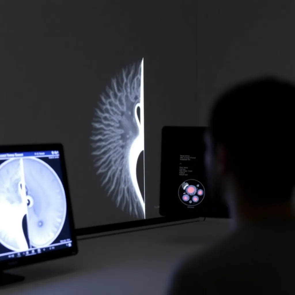
Cervical cancer is the fourth most common cancer among women globally, posing a significant threat to women’s health. Early detection and accurate diagnosis are critical for improving treatment outcomes and reducing mortality rates. Magnetic resonance imaging (MRI) has emerged as the preferred imaging modality for diagnosing, staging, and evaluating cervical cancer, as it provides detailed anatomical information and enhanced contrast between the tumor and surrounding tissues.
Revolutionizing Cervical Cancer Detection with AI
Recognizing the urgent need for automated and efficient cervical cancer detection methods, a team of researchers has developed a novel AI model called SwinUNeCCt. This cutting-edge system leverages deep learning techniques to analyze MRI scans and accurately identify and characterize cervical cancer lesions.
The SwinUNeCCt Approach
The SwinUNeCCt model incorporates a unique bidirectional hash-based agent multi-head self-attention mechanism, which allows the model to better understand the complex patterns and structures of cervical cancer in MRI scans. This mechanism enables the model to effectively interact between local and global features, enhancing its ability to accurately pinpoint and delineate cervical cancer lesions.

Additionally, the SwinUNeCCt model employs a reduced computational complexity self-attention mechanism, making it more efficient and practical for real-world clinical applications. The model also includes a tri-branch inception module that efficiently extracts local features, such as lesion edges and textures, further improving its performance in identifying cervical cancer lesions.
Validating the SwinUNeCCt’s Effectiveness
The researchers thoroughly evaluated the SwinUNeCCt model using a carefully annotated MRI dataset, which included imaging data from 122 cervical cancer patients, each with 100 pelvic dynamic contrast-enhanced MRI scans. The results were impressive, with the SwinUNeCCt model outperforming several state-of-the-art 3D medical segmentation models across multiple key metrics, including the 95th percentile Hausdorff distance (95HD), intersection over union (IoU), and Dice similarity coefficient (DSC).

Fig. 2
Notably, the SwinUNeCCt model achieved a 95HD of 6.25, an IoU of 0.669, and a DSC of 0.802, all of which are the best results among the compared models. This extraordinary performance demonstrates the model’s ability to accurately identify and delineate cervical cancer lesions in MRI scans, paving the way for more personalized and effective treatment strategies.
Unlocking the Potential of Multi-Task Learning
The researchers also explored the impact of joint training on the SwinUNeCCt model’s performance. By incorporating both cervical cancer degree of differentiation and pathological type classification tasks, the model was able to further enhance its semantic segmentation capabilities, highlighting the benefits of multi-task learning for medical image analysis.

Algorithm 1
The ability to simultaneously assess the degree of differentiation and pathological type of cervical cancer lesions can provide valuable insights to clinicians, guiding the development of personalized treatment plans and improving patient outcomes.
Towards Widespread Clinical Adoption
While the SwinUNeCCt model has demonstrated impressive results in the research setting, the researchers acknowledge the need to address several challenges before widespread clinical adoption. These include improving the model’s robustness to noise and image artifacts, as well as exploring strategies to reduce the reliance on large-scale annotated datasets, such as semi-supervised or self-supervised learning techniques.
By addressing these challenges, the SwinUNeCCt model has the potential to revolutionize the way cervical cancer is diagnosed and managed, ultimately improving patient care and outcomes. As the researchers continue to refine and validate the model, the future looks promising for this innovative AI-powered approach to cervical cancer detection and analysis.
Author credit: This article is based on research by Chongshuang Yang, Zhuoyi Tan, YiJie Wang, Ran Bi, Tianliang Shi, Jing Yang, Chao Huang, Peng Jiang, Xiangyang Fu.
For More Related Articles Click Here