Researchers have discovered that college students with depressive states exhibit impaired cognitive control and reduced neural activity when asked to complete a semantic inhibition task. Using a combination of electroencephalography (EEG) and pupillometry, the study sheds light on the neural underpinnings of this deficit, which may contribute to common symptoms of depression, such as rumination and anhedonia. The findings suggest that individuals with depression have difficulty suppressing irrelevant thoughts and processing emotional information, potentially leading to a persistent negative thought cycle. This research provides valuable insights into the neurological mechanisms underlying depression and could have important clinical applications. Depression, Cognitive control, Electroencephalography
Uncovering the Brain’s Struggle with Depression
Depression is a prevalent mental health issue among college students, with around 30% of students experiencing some degree of depressive symptoms. This debilitating condition has been linked to deficits in executive control and inhibitory functions, which can contribute to common symptoms like rumination and anhedonia.
Putting the Brain to the Test
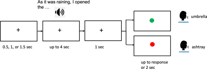
In a recent study, researchers investigated the cognitive and neurophysiological differences between college students with depressive states and their non-depressed peers. The participants were asked to perform a modified version of the Hayling Sentence Completion Task (HSCT), which assesses semantic inhibition – the ability to suppress a strongly associated word and produce an unrelated word instead.
Revealing the Neural Dynamics of Semantic Inhibition
The study utilized a combination of electroencephalography (EEG) and pupillometry to track the brain’s activity and physiological arousal during the HSCT. The researchers found that the students with depressive states showed a reduced neural response, specifically in the frontal N450 component, which is associated with semantic conflict and inhibition.
Importantly, the reduction in N450 was more pronounced in the left hemisphere for the depressed group, suggesting a functional imbalance in the brain’s inhibition-related networks. This finding aligns with previous research, which has identified a pattern of right-hemisphere hyperactivity and left-hemisphere hypoactivity in individuals with major depression.
The Pupil’s Perspective
The pupillometry data revealed an interesting contrast between the two groups. While the control group showed similar pupil dilation for both the initiation and inhibition conditions, the depressed group exhibited greater pupil dilation during the inhibition trials. This suggests that individuals with depression exert more cognitive effort or experience higher arousal when attempting to suppress the prepotent semantic responses.
Connecting the Dots: Symptoms and Neural Activity
The study also explored the connections between the neurophysiological measures and self-reported symptoms of depression. The researchers found that the reduced frontal N450 activity during inhibition trials was associated with higher levels of rumination and anhedonia (a decrease in the ability to experience pleasure) in the participants.
This indicates that the impaired semantic inhibition observed in the depressed group may contribute to the persistence of negative thoughts and the diminished ability to experience positive emotions, which are hallmarks of depression.
Implications and Future Directions
The findings of this study provide valuable insights into the neural underpinnings of cognitive deficits in depression. By combining EEG and pupillometry, the researchers were able to uncover the complex interplay between brain dynamics, physiological arousal, and the specific symptoms experienced by individuals with depressive states.
These insights could have important clinical applications, as they suggest that interventions targeting the improvement of semantic inhibition and the normalization of brain activity patterns may be beneficial for individuals struggling with depression. Future research in this area could further explore the potential for using neurophysiological markers as diagnostic or treatment-monitoring tools for depression.
Author credit: This article is based on research by D. Jan, J. López-Pigüi, Iván Padrón, M. de Vega.
For More Related Articles Click Here