Researchers have developed a groundbreaking device that allows them to simultaneously record electrical signals and observe calcium activity in the hippocampus of mice, a crucial brain region involved in memory and spatial awareness. This innovative soft microelectrode array, integrated into a cranial window, provides a unique window into the inner workings of the hippocampus, enabling scientists to study its complex neuronal networks in unprecedented detail. By leveraging the transparency of the device, researchers can perform high-speed two-photon imaging of calcium signals in the hippocampus while also recording electrophysiological data, including the characteristic sharp wave-ripples and neural oscillations. This groundbreaking approach holds great promise for advancing our understanding of the hippocampus and its role in memory formation, spatial navigation, and neurological disorders.
Revealing the Hippocampus’s Secrets
The hippocampus is a crucial brain region that plays a central role in the formation, consolidation, and recall of memories, as well as in microscopy’>two-photon imaging with electrophysiological recordings in awake, behaving animals. This powerful combination allows researchers to directly observe the activity of individual neurons and neural networks within the hippocampus, while simultaneously recording the electrical signals that underlie their function.
The Soft, Transparent Microelectrode Array
In this groundbreaking study, the researchers developed a unique soft, transparent microelectrode (STM) device that can be implanted directly on the surface of the hippocampus. This innovative device is designed to be highly flexible and responsive, with a Young’s modulus that gradually decreases from the gigapascal (GPa) range to the megapascal (MPa) range as it approaches body temperature. This softening property helps to minimize the immune response and improve the long-term stability of the implant.

Chronic Hippocampal Recordings and Imaging
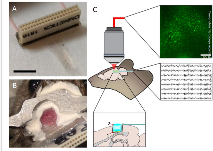
The STM device was incorporated into a cranial window, allowing the researchers to perform high-speed, two-photon calcium imaging of the hippocampal cell’>GCaMP6f-labeled pyramidal cells in the CA1 region of the hippocampus. Remarkably, this optical access was maintained for up to six months, enabling the researchers to study the activity of these neurons over an extended period.

Minimizing the Immune Response
The researchers also examined the long-term immune response to the implanted device through immunohistochemical analysis. They found that while the implantation surgery itself did cause some cellular changes and increased astrocyte activity, the addition of the STM device did not significantly exacerbate these effects. This suggests that the softening properties of the device helped to minimize the immune response and maintain the quality of the recordings over time.
Unlocking the Hippocampus’s Secrets
This innovative study demonstrates the tremendous potential of the soft, transparent microelectrode array for advancing our understanding of the hippocampus. By enabling simultaneous electrophysiological recordings and two-photon imaging, this device provides an unprecedented window into the complex neuronal networks and signaling dynamics within this crucial brain region.
The ability to monitor hippocampal activity over extended periods, while also observing the underlying cellular and molecular changes, opens up new avenues for research into a wide range of neurological processes and disorders, from epilepsy and Click Here