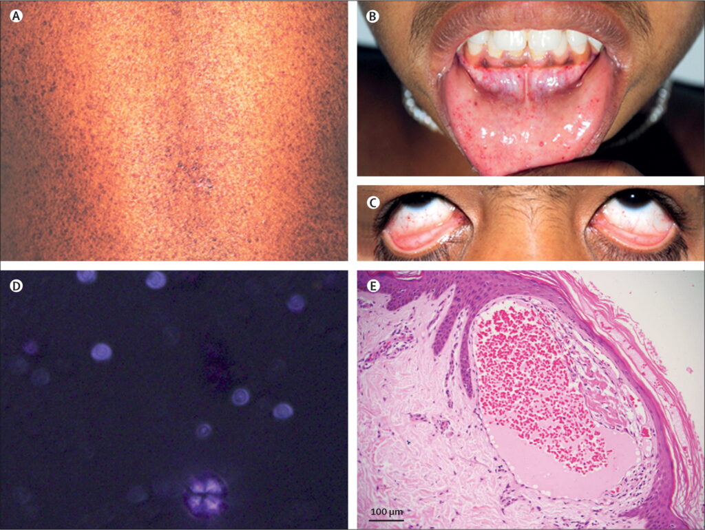
Fabry disease, a rare genetic disorder, can have widespread effects on the body, including the eyes. In this study, researchers uncovered unique insights into how Fabry disease impacts the retinal microvasculature, arterial stiffness, and blood flow in the central retinal artery. By leveraging advanced imaging techniques like optical coherence tomography angiography (OCTA) and color Doppler sonography, the team found that Fabry patients exhibited decreased retinal vessel density, increased arterial stiffness, and elevated resistance in the central retinal artery – even before the onset of overt eye symptoms. These findings shed light on the subtle yet significant ocular changes underlying Fabry disease and highlight the potential of non-invasive biomarkers for early detection and monitoring of this complex condition. Fabry disease is an inherited disorder caused by a deficiency of the enzyme alpha-galactosidase A, leading to the accumulation of glycosphingolipids in various tissues.
Tracking the Earliest Signs of Ocular Involvement
Fabry disease can manifest a wide range of symptoms, including several that affect the eyes. Previous studies have reported corneal opacity, lens changes, and vascular abnormalities in the retina and conjunctiva of Fabry patients. However, these visible eye problems often arise later in the disease progression, making it crucial to identify earlier, subclinical signs of ocular involvement.
In this study, the researchers employed two powerful imaging techniques – OCTA and color Doppler sonography – to delve deeper into the ocular changes occurring in the early stages of Fabry disease. OCTA allowed them to quantify the density of blood vessels in different layers of the retina, while color Doppler provided insights into the blood flow dynamics in the central retinal artery.
Reduced Retinal Vessel Density and Increased Arterial Resistance
The analysis revealed that Fabry patients exhibited a significant decrease in the vessel density of the superficial, deep, and choriocapillaris layers of the retina compared to healthy controls. Notably, the choriocapillaris – the dense network of capillaries that nourish the outer retina – showed the most pronounced reduction in vessel density.
Furthermore, the researchers found that the resistive index of the central retinal artery, a measure of vascular resistance, was notably higher in Fabry patients than in the control group. This suggests that the blood flow in the retinal arteries of Fabry patients is more restricted, likely due to the accumulation of glycosphingolipids in the vessel walls.
Increased Arterial Stiffness: A Systemic Complication
In addition to the ocular changes, the study also revealed that Fabry patients exhibited increased arterial stiffness, as measured by aortic pulse wave velocity. This systemic vascular complication has been well-documented in Fabry disease and is known to contribute to the increased risk of cardiovascular events in these patients.
Implications for Early Diagnosis and Monitoring
The findings of this study underscore the importance of early detection and continuous monitoring of ocular changes in Fabry disease. The observed reductions in retinal vessel density and the elevated resistance in the central retinal artery may serve as non-invasive biomarkers to identify subclinical ocular involvement and track the progression of the disease.
Furthermore, the insights gained from this research can aid in the development of targeted treatment strategies and the assessment of therapeutic responses, particularly for emerging therapies like enzyme replacement therapy. By closely monitoring these ocular parameters, clinicians can gain a more comprehensive understanding of the disease’s impact and tailor interventions accordingly.
A Multifaceted Approach to Fabry Disease Management
Fabry disease is a complex, multisystem disorder that requires a multifaceted approach to management. This study highlights the value of incorporating advanced imaging techniques, such as OCTA and color Doppler sonography, into the clinical assessment of Fabry patients. By gaining a deeper understanding of the subtle ocular changes and systemic vascular complications, healthcare providers can implement more effective strategies for early intervention, disease monitoring, and personalized treatment plans.
As research in this field continues to evolve, the integration of these innovative imaging tools, coupled with a holistic understanding of Fabry disease, will be crucial in improving the quality of life and long-term outcomes for individuals affected by this rare, yet debilitating, genetic condition.
Author credit: This article is based on research by Michele Rinaldi, Flavia Chiosi, Maria Laura Passaro, Francesco Natale, Alessia Riccardo, Luca D’Andrea, Martina Caiazza, Marta Rubino, Emanuele Monda, Gilda Cennamo, Francesco Calabrò, Giuseppe Limongelli, Ciro Costagliola.
For More Related Articles Click Here
