Cataracts, the clouding of the eye’s lens, are a common condition affecting both adults and children. While adult cataracts are often age-related, childhood cataracts can have various underlying causes, including a rare congenital condition called Persistent Hyperplastic Primary Vitreous (PHPV). In this comprehensive study, researchers from South Korea delved into the histopathological characteristics of PHPV and the thickness of the lens capsule in pediatric cataract patients. Their findings shed new light on the mechanisms behind this complex eye disorder and provide valuable insights for clinicians and researchers alike.
Unraveling the Mysteries of Persistent Hyperplastic Primary Vitreous
PHPV is a rare congenital eye anomaly where the hyaloid vessel system, a transient intraocular vascular system formed during eye development, fails to regress after birth. This can lead to various complications, including cataracts, intraocular hemorrhage, and even vision loss. Understanding the pathogenesis of PHPV is crucial for improving treatment and management strategies for affected children.
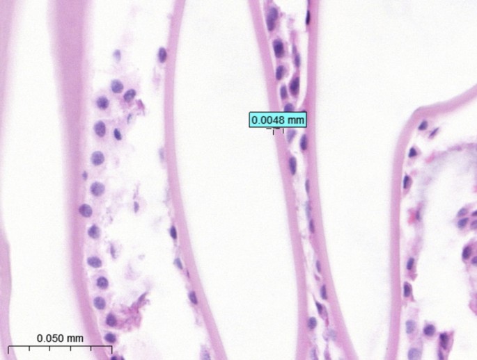
The researchers analyzed lens capsule samples from 116 eyes of 83 Korean pediatric cataract patients, including 13 cases of PHPV. They measured the thickness of the anterior and posterior lens capsules and conducted detailed histopathological and immunohistochemical examinations of the retrolental membranes in PHPV cases.
Lens Capsule Thickness: Unaffected by Age or PHPV
One of the key findings of the study was that the mean thickness of the anterior and posterior lens capsules did not differ significantly based on the patients’ age, gender, or the presence of PHPV. This suggests that the lens capsule, which serves as a basement membrane for lens epithelial cells, is not significantly affected by the delayed eye development often seen in PHPV.

Endothelial-Mesenchymal Transition: A Potential Mechanism in PHPV
The histopathological analysis of the retrolental membranes in PHPV cases revealed an interesting pattern. While some cases showed the presence of endothelium-lined blood vessels, the majority (69%) exhibited a predominance of mesenchymal cells, with or without vascular structures. This finding supports the hypothesis that endothelial-mesenchymal transition (EndMT), a process where endothelial cells acquire a mesenchymal phenotype, may be a key mechanism in the vascular regression seen in PHPV.

Diverse Histological Characteristics of PHPV
The researchers also found that not all PHPV cases exhibited the classic histological features of vascular structures in the retrolental membranes. In some cases, the fibrovascular membrane tissue was too small to be detected through histology, while in others, only mesenchymal cells were present without any visible vascular components. This suggests that the regression of the hyaloid vascular system in PHPV may not always follow a uniform pattern, highlighting the complex and diverse nature of this condition.
Implications for Diagnosis and Treatment
The findings of this study have important implications for the diagnosis and management of pediatric cataracts associated with PHPV. The lack of significant differences in lens capsule thickness between PHPV and non-PHPV cases suggests that this parameter may not be a reliable indicator of PHPV. Additionally, the diverse histological characteristics of PHPV, with varying proportions of vascular structures and mesenchymal cells, underscore the need for a comprehensive clinical and histopathological evaluation to accurately diagnose and manage this condition.
Future Research Directions
This study opens up new avenues for further research into the pathogenesis of PHPV. Exploring the role of Click Here